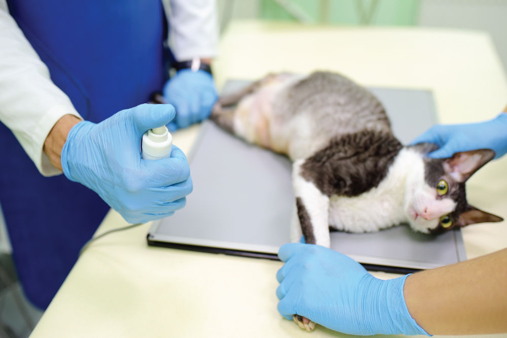
Diagnostic ultrasound, which uses high-frequency sound waves to create real-time images of soft tissue anatomy, is another significant advancement in veterinary medicine. As a safe, painless way to look inside the body, ultrasound can observe blood flow, heartbeats, and gastrointestinal movement as they occur.
Abdominal ultrasound can be used to examine soft tissue structures such as the liver, gall bladder, spleen, kidneys, bladder, prostate, uterus, ovaries, adrenal glands, stomach, and intestines.
Thoracic (chest) ultrasound can be used to uncover abnormalities of the heart and surrounding structures, such as increased fluid, enlarged lymph nodes, and tumors.
If your pet has symptoms such as excessive vomiting, infrequent urination, unexplained weight loss, chronic infections, or has received abnormal blood work results, we may request to perform an ultrasound. It is also used as part of a presurgical screening, to diagnose pregnancy, to determine staging in cancer patients, and as a baseline by which to compare results of future visits (e.g., geriatric patients.)

X-rays (also known as radiographs) are utilized to evaluate organs (heart, lungs, kidneys, abdomen, etc.), soft tissue (such as muscle or tendon), and dense tissue (such as bones). Changes in shape, size, and position of organs can be an indicator of disease. X-rays can also uncover medical issues such as fractured bones, dislocated joints, infection, abnormal bone growths, changes due to metabolic bone disease, and conditions such as arthritis.
Multiple images taken from different angles provide a comprehensive look at an area of interest. Our experienced veterinarians and licensed technicians are highly skilled at obtaining and interpreting these images.
Dugan’s Veterinary Hospital offers digital x-rays that produce immediate results, reduce exposure to radiation and require less table time for your pets versus the traditional film method. Digital x-rays are also ideal for uncovering tooth decay and periodontal disease that can be hidden from the naked eye and thus missed during a dental exam.

For pet ultrasound in Aurora, CO, turn to Dugan's Veterinary Hospital. Our advanced diagnostic equipment can help find hidden signs of illness, injury, or disease in your pet, allowing us to provide the best course of treatment to return your best friend to health. To find out more about ultrasound procedures or to schedule your pet’s appointment, please call now.